
Three Tests That Look Inside the Body in Three Different Ways
After a car accident, imaging is often one of the first formal medical evaluations a patient receives. An emergency room may order an X-ray. A specialist may follow up with a CT scan. A few weeks later, an MRI may be requested.
To most patients, these three tests can feel interchangeable. They are not. Each uses distinct physical principles, exhibits different structures, poses different risks, and addresses distinct clinical questions. Understanding what each test actually does can help an injured individual make better sense of the care they are receiving and the reports that result from it.
X-Ray: The Workhorse for Bones
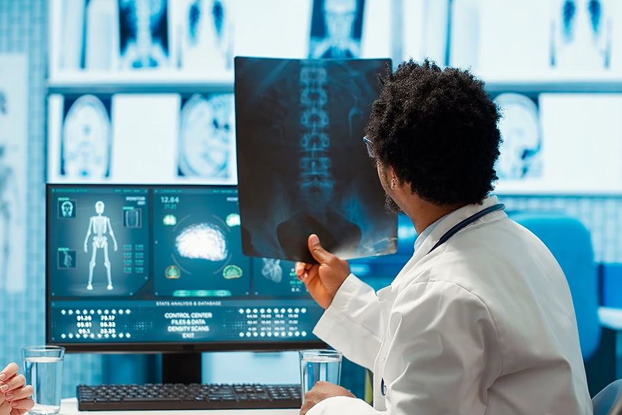
The X-ray is the oldest and most widely used form of medical imaging.
According to the National Institute of Biomedical Imaging and Bioengineering (NIBIB), an institute of the National Institutes of Health, X-rays are a form of electromagnetic radiation, similar to visible light. Unlike light, X-rays have higher energy and can pass through most objects, including the body. Medical X-rays are used to generate images of tissues and structures inside the body.
How It Works
According to NIBIB, when X-rays pass through the body, they reach an X-ray detector on the other side of the patient, forming an image that represents the “shadows” of the objects inside the body. Dense structures, such as bone, absorb X-rays readily and appear bright white on the image. Soft tissues, such as muscle and fat, absorb radiation less readily and appear in shades of gray.
What It Shows
X-rays excel at imaging bone. According to NIBIB, X-ray scans can diagnose conditions including bone cancer, infections, and blocked blood vessels. In the context of a car accident, an X-ray is most useful for identifying fractures, dislocations, and obvious bony abnormalities.
What It Misses
X-rays show bone clearly. They show soft tissue poorly. A patient leaving the emergency room with a normal X-ray has been told that no fracture has been identified. They have not been told anything definitive about the muscles, ligaments, tendons, discs, or nerves that may have been damaged in the collision.
Radiation Considerations
According to the NIBIB fact sheet, when used appropriately, the diagnostic benefits of X-ray scans significantly outweigh the risks. For conventional X-rays, the amount of radiation delivered to a patient is extremely small.
CT Scan: Cross-Sectional Detail of Bone and More
The CT scan, sometimes called a CAT scan, is essentially a sophisticated form of X-ray imaging.
According to NIBIB, the term computed tomography, or CT, refers to a computerized X-ray imaging procedure in which a narrow beam of X-rays is aimed at a patient and quickly rotated around the body, producing signals that are processed by the machine’s computer to generate cross-sectional images, or slices. These slices are called tomographic images and can give a clinician more detailed information than conventional X-rays.
How It Works
According to NIBIB, unlike a conventional X-ray, which uses a fixed X-ray tube, a CT scanner uses a motorized X-ray source that rotates around the circular opening of a donut-shaped structure called a gantry. During a CT scan, the patient lies on a bed that slowly moves through the gantry while the X-ray tube rotates around the patient, shooting narrow beams of X-rays through the body. Once a series of successive slices is collected by the machine’s computer, they can be digitally stacked to form a three-dimensional image of the patient.
What It Shows
According to NIBIB, a CT scan is particularly useful for imaging bone fractures, joints, cartilage, or tendons, as it usually provides more detail than a conventional X-ray. CT can also be used to image the head in order to locate injuries, tumors, clots leading to stroke, hemorrhage, and other conditions.
In the context of a car accident, CT is the imaging test most commonly used in emergency settings when a more detailed view of the bones is needed than an X-ray can provide. It is particularly valuable for identifying skull fractures, intracranial bleeding, complex fractures, and certain types of internal injury.
According to StatPearls, a clinical reference published through NCBI Bookshelf, the advantage of these tomographic images over conventional X-rays is that they provide detailed information about a specific area in cross-section, eliminating image superimposition and offering a tremendous advantage over plain films.
What It Misses
CT excels at showing bone and identifying acute findings such as bleeding. It is less detailed than MRI for soft tissue evaluation. Subtle ligament injuries, early-stage disc herniations, and many rotator cuff or labral injuries may not be apparent on CT.
Radiation Considerations
CT involves significantly more radiation exposure than a single X-ray because the technique captures multiple images. According to NIBIB, all X-rays produce ionizing radiation, which can cause biological effects in the human body. Modern CT protocols are designed to minimize this exposure, and clinical use is appropriate when the diagnostic benefit warrants it.
MRI: The Gold Standard for Soft Tissue
MRI uses an entirely different physical principle than X-ray or CT. It does not use radiation at all.
According to NIBIB, MRIs employ powerful magnets that produce a strong magnetic field that forces protons in the body to align with that field. When a radiofrequency current is then pulsed through the patient, the protons are stimulated and spin out of equilibrium, straining against the pull of the magnetic field. When the radiofrequency field is turned off, the MRI sensors are able to detect the energy released as the protons realign with the magnetic field. The time it takes for the protons to realign with the magnetic field, as well as the amount of energy released, changes depending on the environment and the chemical nature of the molecules. Physicians are able to tell the difference between various types of tissues based on these magnetic properties.
How It Works
According to StatPearls, the components of the MRI machine include a set of primary magnets, three gradient coils, shim coils, and an integrated radiofrequency coil. The strength of the magnetic field is measured in Tesla. Clinical MRI is usually performed at 1.5 to 3 Tesla.
What It Shows
MRI is the most powerful imaging modality currently available for evaluating soft tissue. It can show muscles, ligaments, tendons, intervertebral discs, the brain and spinal cord, nerves, the joints in detail, and the internal structures of the abdomen and pelvis with a level of contrast that no other imaging test can match.
In the context of a car accident, MRI is the test most commonly ordered when:
- A herniated disc is suspected
- Rotator cuff or labral injury of the shoulder is being evaluated
- ACL, MCL, or meniscal injury of the knee is being assessed
- A concussion has produced lingering symptoms requiring further evaluation
- Soft tissue injury that did not appear on X-ray is suspected
What It Misses
MRI is less detailed than CT for acute bony detail and certain types of acute bleeding. It is also more expensive, takes longer to perform (often 30 to 60 minutes), and requires the patient to lie still inside an enclosed magnetic tube, which can be difficult for patients with claustrophobia or significant pain.
Safety Considerations
MRI does not use ionizing radiation. However, the magnetic field is exceptionally strong. According to NIBIB, the magnetic field extends beyond the machine and exerts very powerful forces on objects of iron, some steels, and other magnetizable objects. Patients should notify their physicians of any implants, metal fragments, or medical devices before undergoing MRI.
People with certain implants, including pacemakers, vagus nerve stimulators, implantable cardioverter defibrillators, insulin pumps, cochlear implants, deep brain stimulators, and certain other devices, may not be able to undergo standard MRI safely. NIBIB notes that loud noise, sometimes reaching up to 120 decibels in certain MRI procedures, is also a consideration.
How These Tests Compare in Practice
| Comparison | X-Ray | CT | MRI |
|---|---|---|---|
| Best for | Bone fractures and dislocations | Detailed bony anatomy, acute bleeding, and complex fractures | Soft tissue, discs, ligaments, brain, and spinal cord |
| Uses radiation | Yes, low dose | Yes, higher dose | No |
| Time required | Minutes | Minutes | 30–60 minutes |
| Soft tissue detail | Limited | Moderate | Excellent |
| Cost | Lowest | Moderate | Highest |
| Availability in ER | Universal | Common | Less common |
What Imaging Reports Add to the Clinical Record
Each of these imaging studies generates a formal report written by a radiologist. The report includes a description of the findings observed in the images and an interpretation of their meaning. Together, the imaging studies and their reports form part of the complete clinical record of an injury.
For accident victims whose recovery may extend over weeks or months, this record accumulates. An initial X-ray. A later CT. A subsequent MRI. Each becomes part of the timeline documenting the body’s state at specific points after the collision. Comparison across these studies, by treating clinicians as care progresses, often reveals more than any single study viewed in isolation.
How AP Healthcare Can Help
Imaging studies are rarely a single event after a car accident. A patient may undergo initial imaging in the emergency department, follow-up imaging weeks later as ordered by a specialist, and additional studies as treatment progresses. Each study generates a report. Each report becomes part of a complete clinical record that someone has to track, organize, and ensure is accessible when needed.
AP Healthcare serves as a concierge for post-accident care coordination. We are not a medical provider and do not offer medical advice; those decisions remain between the patient and their healthcare team. We do not determine treatment, imaging, or provider choices; those decisions are made by the patient and their healthcare team.
What we do is manage the logistics that surround care. We help connect injured individuals with experienced providers, assist with scheduling for imaging and follow up appointments, arrange transportation when getting to appointments is a challenge, and provide translation services when language is a barrier. We also assist with the collection and organization of imaging reports and medical records throughout the treatment process, so that the full clinical picture remains accessible from the first study through the final follow up.
To learn more, visit aphealthcare.org or call (404) 850-9600.
This article is for informational purposes only and does not constitute medical or legal advice. Please consult a qualified healthcare provider for guidance specific to your situation.
Sources:
- National Institute of Biomedical Imaging and Bioengineering (NIBIB), National Institutes of Health — Medical X-rays — fact sheet updated June 2022
- National Institute of Biomedical Imaging and Bioengineering (NIBIB), National Institutes of Health — Computed Tomography (CT) — fact sheet updated June 2022
- National Institute of Biomedical Imaging and Bioengineering (NIBIB), National Institutes of Health — Magnetic Resonance Imaging (MRI)
- National Cancer Institute (NCI), National Institutes of Health — Computed Tomography (CT) Scans and Cancer Fact Sheet
- NCBI Bookshelf / StatPearls — CT Scan — Patel PR, De Jesus O
- NCBI Bookshelf / StatPearls — CT-scan Image Production Procedures — Hermena S, Young M
- NCBI Bookshelf / StatPearls — Magnetic Resonance Imaging Physics
- NCBI Bookshelf / StatPearls — Lumbar Disc Herniation — Al Qaraghli MI, De Jesus O